
A CT (Computed Tomography) scan provides vital diagnostic information by creating highly detailed, three-dimensional images of internal organs. This non-invasive imaging technique uses X-ray technology to scan specific areas of the body, enabling the accurate diagnosis of various medical conditions across multiple organ systems.
To schedule your CT scan, contact us by phone at 050-475-1120 or email us at office@rdg-imaging.com. We prioritize high availability and prompt response to every inquiry.
Our team will help coordinate your appointment at a time that is most convenient for you, considering both your medical urgency and personal schedule. Your request will be reviewed together with your referral and any relevant clinical documentation to ensure the exam is tailored precisely to your medical question.
You’ll receive detailed preparation instructions in advance (e.g., fasting, drinking fluids, or medication, if needed) to ensure optimal image quality and accurate results.
Although most imaging centers provide a basic interpretation, many patients seek a second opinion or more thorough and faster reports. This is where RDG’s team of expert radiologists comes in. We offer professional interpretation by highly experienced radiologists with relevant subspecialty training within 24 to 48 hours.
At RDG, we emphasize uncompromising professionalism, complete transparency, and strict privacy protection throughout the entire process.
All interpretations are performed by experienced subspecialized radiologists, adhering to the highest professional standards. The service is available to any patient who has had a CT scan, regardless of where it was performed—making it an ideal solution for those seeking fast, expert-level analysis.
You can contact us by phone or email (050-475-1120 / office@rdg-imaging.com).
Alternatively, you can easily place an order through our secure website:
Once submitted, we will verify image quality and send you a secure payment link. The interpretation will begin immediately upon confirmation. Our system ensures full compliance with data privacy laws. Personal support is available throughout the process.
After receiving your CT scan and medical documentation, the case is reviewed by a subspecialized radiologist. Each image is carefully analyzed, findings are interpreted, and a comprehensive medical report is written. If complex or abnormal findings are present, internal consultation with other radiology experts may be conducted to ensure diagnostic accuracy.
Upon completion, you will receive a detailed report via a secure link sent to both your email and phone (SMS).
You can view the report from any device, including your smartphone. Findings are explained clearly so that every patient can understand their medical meaning. In special cases, you can schedule a follow-up consultation with the interpreting physician.
Results are usually available within 24 to 48 hours after receiving all required materials and completing payment. Every step is carried out under strict data security protocols to protect your privacy and rights.
Our goal is not only to deliver accurate results but also to provide clarity, professional support, and personalized service. We bring modern diagnostic imaging directly to you—with high availability, expert radiologists, and care that adapts to your needs. This is a perfect combination of advanced technology, clinical expertise, and human-centered service without compromise.
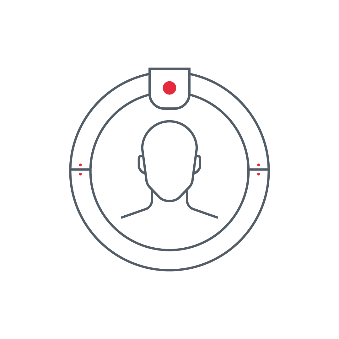
CT head, also known as Brain CT, provides updated and high-quality images of the brain, including brain structure, blood vessels, skull bones, and facial bones. This imaging test may allow the diagnosis of conditions such as infections, ischemia, tumors, fractures, accumulation of fluids, or congenital anomalies. Additionally, head CT scans can include angiography, an examination focusing on the blood vessels of the head and neck, including veins and arteries, efficient for detecting aneurysms, stenosis, or vascular malformations
An advanced and innovative imaging test, the CBCT , produces detailed images of the sinuses and ears with lower radiation dose compared to regular CT scans. Unlike conventional CT or X-ray images taken from different directions, CBCT creates a more detailed three-dimensional image of the four sinuses. This test aids in the diagnosis of various ear, nose, and throat conditions, including growths, polyps, infections, fractures, and cracks in the sinuses and ears, as well as sinusitis

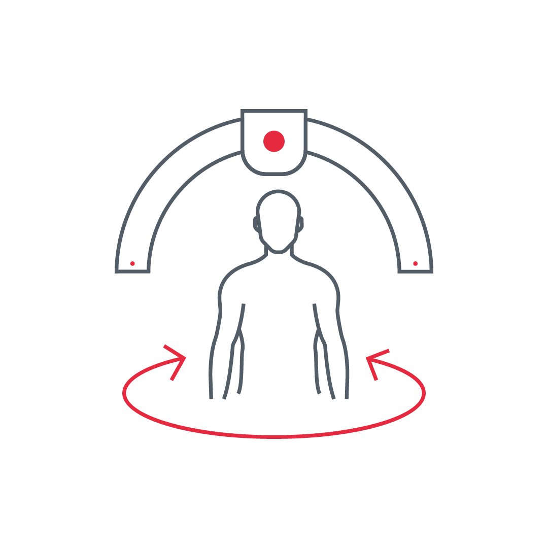
This test covers the area from the base of the skull to the upper chest, including the respiratory passages, lymph nodes, thyroid gland, blood vessels, throat, larynx, and more. It helps diagnose various clinical conditions in these areas, including space occupying lesions, congenital anomalies, inflammatory masses, cysts, inflammation, and vocal cord lesions. Notably, for tumors, CT neck allows not only diagnosis but also helps identify the stage of the disease and plan appropriate treatment, including radiation therapy and tumor removal
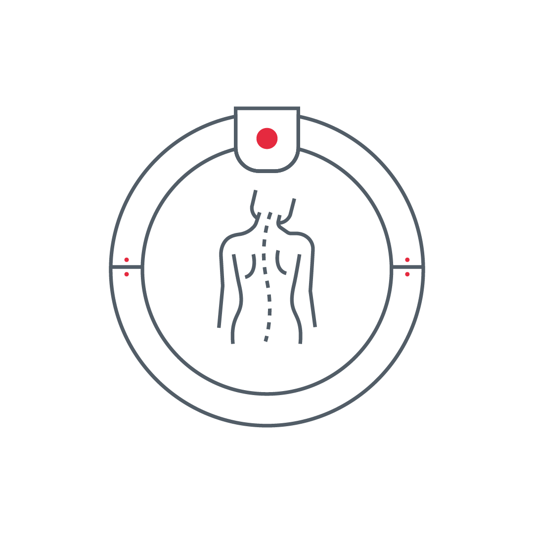
CT spine examinations are divided into three main types: cervical, thoracic, and lumbar. These imaging exams depict, among other things, the spinal cord canal, disc spaces, muscles, and blood vessels in the area. These tests are efficient for identifying the source of pain that may indicate a problem in the spine, including disc rupture, spinal cord stenosis, disc fractures, changes in the vertebrae, infections, and more. Additionally, spinal CT exams are useful for detecting spinal tumors and tracking their spread precisely. Moreover, spinal CT is effective for preparing for invasive treatments in the area, providing an up-to-date image and a detailed assessment of the area before medical procedures
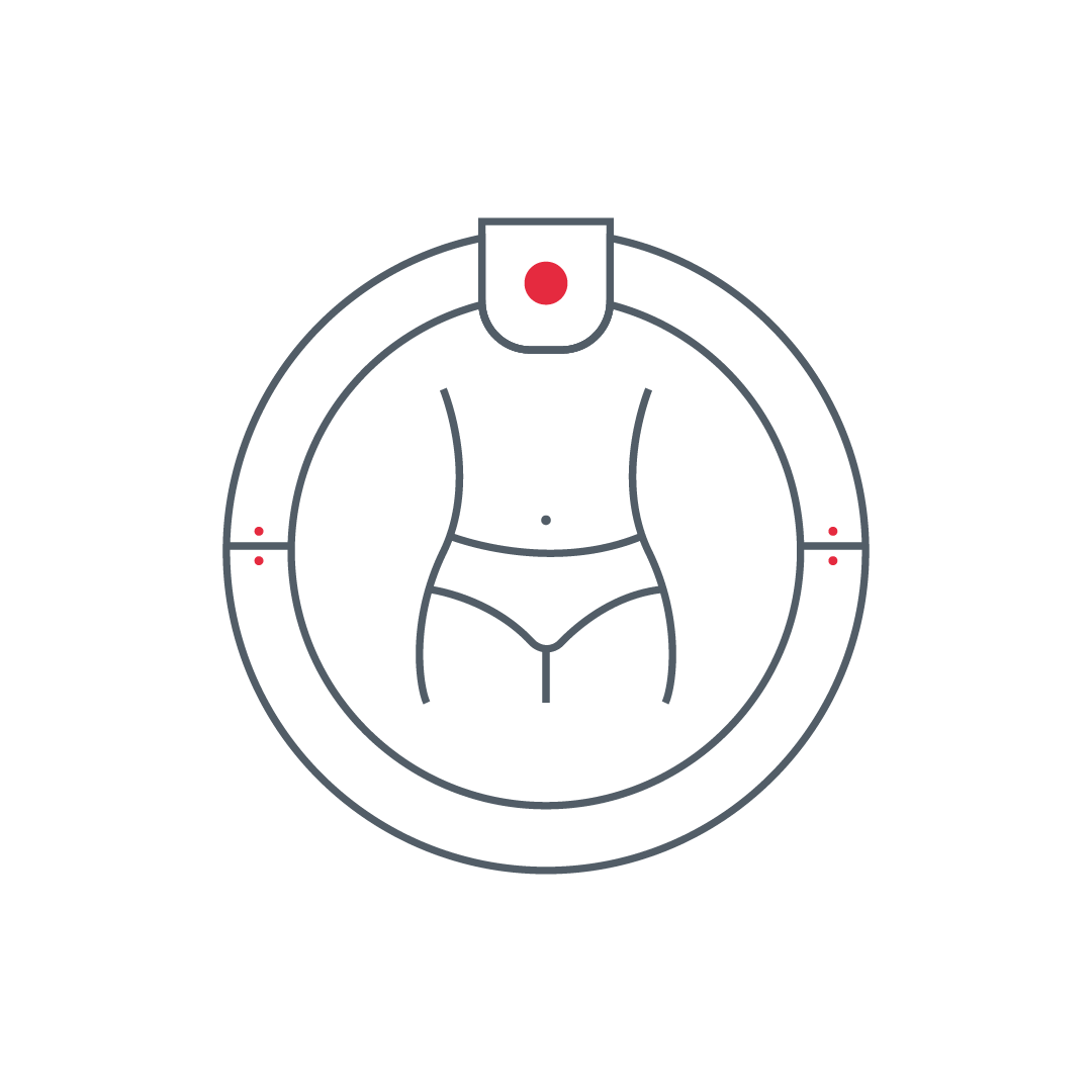
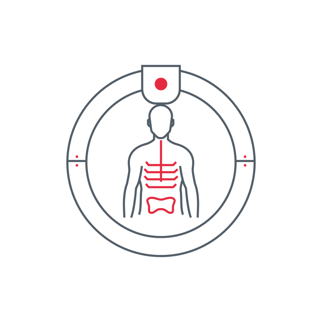
This test generates a detailed image of abdominal organs from different angles, including the digestive system, bones, muscles, and blood vessels. Abdominal CT provides more detailed information compared to abdominal ultrasound or regular X-rays. This test allows the identification of conditions such as tumors, inflammation, infections, bowel obstructions, kidney and gallstone issues, and other diseases in the abdominal and pelvic areas
An examination demonstrating internal organs in the chest area, including lungs, blood vessels, heart, arteries and veins, muscles, and bones. Chest CT helps diagnose lung diseases, growths, and abnormalities in the area, infections, inflammation, respiratory tract disorders, and more. Furthermore, chest CTs can include specific CT exams, such as angiography of the pulmonary arteries (to detect blood clots and lung infarctions), aorta angiography (to identify aneurysms in the arterial system), and CT of the coronary arteries
An examination highlighting soft tissues, bones, and blood vessels. It is effective for diagnosing various medical conditions, including fractures, tumors, infections, sports injuries, joint injuries, inflammation, and neurological conditions

PET-CT combines computed tomography (CT) and functional imaging (PET), accurately visualizing physiological processes alongside detailed anatomical structures. This test is typically used to detect and diagnose cancers, disease staging, infections, inflammations, and neurological or cardiac disorders. Additionally, PET-CT is valuable for monitoring responses to oncology treatments and assessing disease spread throughout the body. At RDG Imaging, we provide unique and detailed reporting for PET-CT exams, which includes: A full radiological report of the findings in the whole-body CT scan interpreted by an experienced radiologist. A detailed evaluation of the nuclear imaging (PET) results performed by a specialist in nuclear medicine, as well as interdisciplinary exchange between the two specialists, combining their impressions to provide a comprehensive and accurate assessment of the patient’s condition. Our unique approach ensures that patients receive extensive and detailed information about their disease status, integrating precise anatomical interpretation with functional imaging analysis for optimal treatment planning.

Cardiac CT provides a highly detailed and accurate visualization of the heart and coronary blood vessels. This test is typically performed to diagnose cardiovascular diseases, such as coronary artery stenosis or blockages, assess post-catheterization or surgical outcomes, and detect congenital anomalies. Cardiac CT is an advanced diagnostic tool that enables precise treatment planning, including valve assessment, calcium buildup detection in blood vessels, and non-invasive evaluation of heart function. A significant advantage of this test is its ability to provide high-resolution imaging essential for accurate medical decision-making. Additionally, Cardiac CT serves as a valuable tool for monitoring individuals at risk of heart disease, allowing for early detection of atherosclerotic changes even before clinical symptoms appear.