How to Schedule an Ultrasound Appointment with RDG (Clinic or At-Home)?
Booking an ultrasound appointment with RDG—whether at your home or one of our medical clinics—is simple, quick, and personalized. We offer several convenient contact methods:
- By phone: Call us at 050-475-1120 to speak with one of our representatives who will help you schedule your exam at a time that works best for you. You can also leave us your details on our homepage, and we’ll get back to you promptly.
- By email: Send a message to office@rdg-imaging.com with the following details:
- Full name
- Phone number
- Type of service requested: At-home or in-clinic ultrasound
- Your location (for home visits)
- Referral or exam details, if available
We will contact you with available time slots and relevant instructions. We are committed to providing prompt, courteous, and personalized responses. In most cases, we can offer an appointment within a short time frame based on team availability and your location.
Appointments are adapted to your schedule—including evenings, weekends, and urgent requests. If special preparation is needed (e.g., fasting, drinking water, or specific clothing), you’ll receive clear instructions in advance.
RDG Ultrasound – At Home or in Clinic
RDG’s ultrasound services offer advanced, accurate, and accessible diagnostic imaging tailored to your individual needs. You can choose to:
- Have the scan performed in one of our medical clinics with state-of-the-art equipment
- Or receive the exam in the comfort of your home with portable modern devices
In-clinic exams are conducted by an experienced radiologist. A certified technician performs at-home exams. In both cases, the service is pre-scheduled to align with your availability and medical needs.
All ultrasound scans are interpreted by a board-certified radiologist with subspecialty expertise.
Results are delivered promptly, accompanied by professional support, clear explanations, and the opportunity to speak directly with the interpreting radiologist.
Who Should Choose At-Home Ultrasound?
- Patients with limited mobility
- Post-surgical patients
- Elderly individuals
- Busy professionals
- Those avoiding exposure to medical facilities or long wait times
Who Should Choose In-Clinic Ultrasound?
- Patients requiring urgent or high-precision imaging in a clinical setting
- Those who prefer a structured medical environment with advanced technology
- Individuals with complex medical histories need closer supervision
- Patients undergoing routine follow-up exams
- Anyone referred by an HMO, specialist, or hospital wishing for quick and private service
- Patients who prefer the scan to be performed by a radiologist rather than a technician
- Anyone seeking professional, accurate diagnostics and expert interpretation
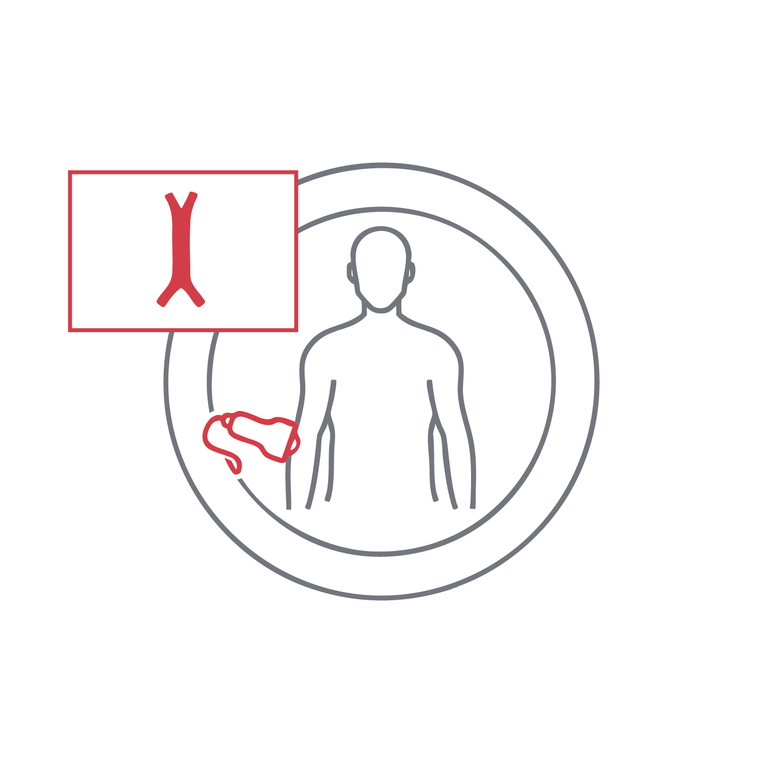
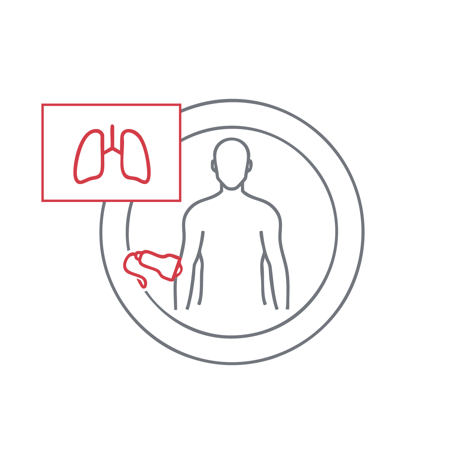
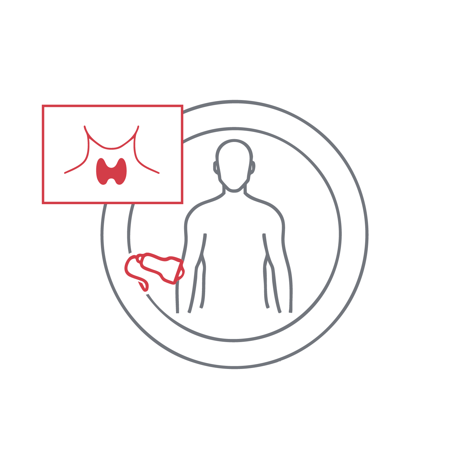
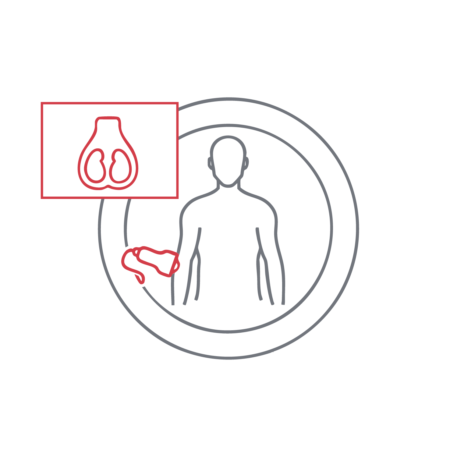
Common studies include abdominal, renal, bladder, thyroid, testicular, neck, and limb vein ultrasounds. Follow-up or focused scans can be performed based on physician referral.
Portable devices offer high-resolution imaging that meets the highest clinical standards. The exam is done privately, in a calm and proper environment, with no compromise on quality or professionalism. Flexible scheduling is available, including urgent requests. Each study is interpreted in the full context of the patient’s medical history and clinical background.
How Is the Ultrasound Performed?
At Home:
After scheduling, a certified technician visits your home with mobile equipment. The scan is performed on your bed or another comfortable surface in a respectful and quiet setting. The procedure follows the same clinical standards as in a medical center.
In Clinic:
You will check in briefly before meeting a radiologist who performs the scan using gel and a transducer over the examined area. The procedure is non-invasive, painless, and takes about 10–30 minutes. The radiologist reviews the images in real-time and records findings.